- Home
- For Researchers & Academics
- Core Facilities
Histology and Microimaging Core (HMC)

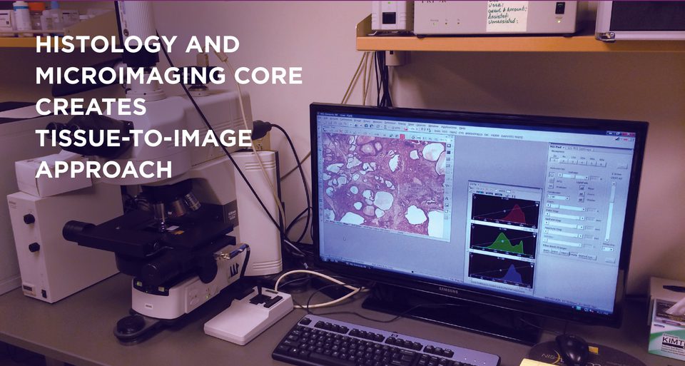
The HMC facility at MWRI provides investigators with high quality, rapid tissue processing and imaging services. The core is designed with a “tissue to image” approach that allows investigators to request services, drop off samples, and receive interpretation, images, or image analyses of their processed samples according to their needs. The histology core is directed by Dr. Carlos Castro.
The HMC, which occupies 730 square feet of laboratory space on the 7th floor of MWRI, houses several integral pieces of equipment, including:
- Leica CM1850UV cryostat for frozen tissue
- Thermo embedding station
- Thermo, Microm, and Leica microtomes
- Complete grossing station
- Manual staining station capable of performing routine, special, and in situ staining procedures
- Leica BondMax Immunostaining System for immunohistochemistry
- Leica LMD 7000 Laser Capture Microdissection (LCM) System
The core operates a routine Sakura Tissue-Tek VIP conventional tissue processor, as well as a Milestone RHS microwave tissue processor, which has several advantages including faster processing times and ease of use when compared to conventional alcohol-based processing.
In combination with special alcohol-based fixatives, the microwave-processed tissue allows the investigator to retrieve macromolecules (DNA, RNA, protein) from the paraffin block at similar quality and quantity compared to bulk flash frozen tissue.
The HMC also houses a full-service microimaging and analysis suite that features a Nikon Eclipse Ni-E, fully motorized upright microscope for capturing high quality and high-resolution bright field, DIC, and fluorescence images.
The core also features a confocal unit, directed by Judy Yanowitz, PhD. It includes a Nikon A1 advanced confocal system with resonant scanner for live-imaging and spectral detection, allowing detections of up to 32 color channels with Volocity 3D/4D imaging and deconvolution software, a Leica DMIRE2 laser confocal microscope, and a Nikon Eclipse TE2000-E spinning disk confocal microscope. All three confocal microscopes are equipped for live cell imaging.
A separate computer workstation offers Photoshop, MetaMorph and Nikon Elements analysis software for more extensive post acquisition image analyses.
Core Services
Services offered:
- Process fresh and fixed tissue specimens (grossing, tissue processing, embedding, sectioning)
- Microwave tissue processing (retrieve quality RNA/DNA from specially fixed paraffin-embedded blocks) and conventional alcohol-based tissue processing
- Frozen sections (performed by investigator, tutorials by core)
- Routine, special, and immunohistochemical stains, including antibody work-up
- Laser capture microdissection
- Imaging (brightfield, epifluorescence, and DIC done in conjunction with core staff) and image analysis on the Nikon Eclipse Ni-E fully motorized imaging platform using Nikon Elements AR software for acquisition and analysis (measuring objects/region of interest, quantifying IHC staining or other staining etc.)
- Training Sessions: Training sessions are available for the cryostat, LCM, and Scope, as well as consultation for special requests/projects through Dr. Castro and Gabrielle King, MS.
Dr. Castro is available for special consultation and interpretation of tissue-based specimens.
Fee Structure:
Contact core personnel for current pricing fee.
Checklist for Users
- Complete histology request form, or at the Core.
- Drop request form in the lab or e-mail completed histology request form to ccastro@mwri.magee.edu or kingg3@upmc.edu
- Drop off tissue and request in lab.
Scheduling Time
- Cryostat time can be scheduled in Outlook at “MWR Histology Core Leica Cryostat” Service Account.
- Microscope time can be scheduled in Outlook at “mwrnikon90i” Service Account.
- LCM time can be scheduled in Outlook at “mwrlasercapture” Service Account.
- Reservation entries must include full name, lab #, extension, and the grant number to be charged.
- Reservations no longer have a 4-hour time limit. Instead of honoring cancellations prior to 4 hours before the session, we will honor cancellations that occur any time before the beginning of the scheduled session. We also ask that any time modifications to the original scheduled session are made as soon as the session is finalized if needed. For example, someone ending a session earlier than originally scheduled should make that change as soon as they are finished.
Additional Resources
New to the Histology Core? Not sure how this technology can help your research?
The Histology Core provides histology research and microimaging research investigators with the highest quality tissue processing, from fresh tissue to digital image and image analysis.
Links to protocols and related websites:
Location
Histology and Microimaging Core (HMC)
Magee-Womens Research Institute
204 Craft Avenue, Room B724.1
Pittsburgh, PA 15213
P: 412-641-5675
Contact
Core Director
Carlos A Castro, DMD, MD
Senior Histotechnician
Gabrielle King, MS
Be the First to Know
Get the latest research, news, events, and more delivered to your inbox.